
How the same technique helped reveal both a 6th century medical text and an ancient bird feather’s pigment.
October 9, 2019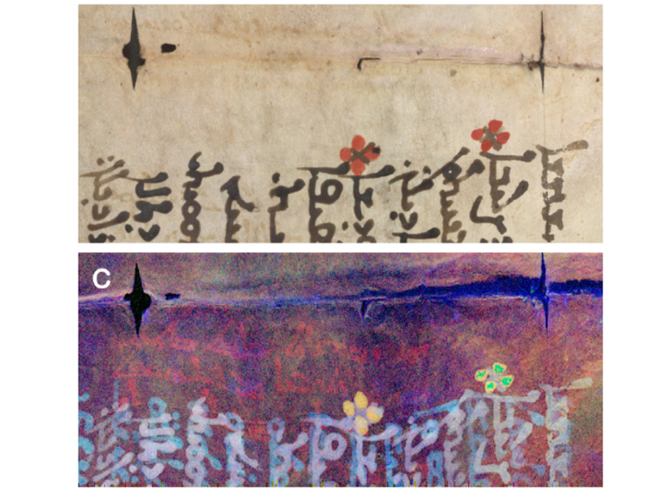
The feathered edges of an 11th century book of hymns fan out as a researcher carefully opens it. The delicate pages, treated with extreme care, hide a secret only revealed with X-ray light. Beneath the hymns lie an even older book of medical care, written by physician and philosopher Galen of Pergamon. The underlying text dates to the 6th century.
It’s the 11th century and parchment is very expensive. A common practice at the time reused older books by scraping their pages clean and writing new material on them. Galen’s book on medicine succumbed to this fate. An X-ray source at the Department of Energy’s SLAC National Accelerator Lab (SLAC) revealed these hidden lines, using a technique that has also revealed the pigment of an ancient bird feather.
The pages of these books, known as palimpsests, were scraped clean and then overwritten with new text. An X-ray technique called X-ray fluorescence at SLAC allows researchers to see the metal traces of the erased text underneath the visible words or images. While X-ray fluorescence is a common technique, the concentrated X-ray beam produced at SLAC allows for greater detail and much faster imaging speed, critical for producing images of whole pages from a palimpsest. The ability to apply a concentrated beam to rapidly image the traces of hidden ink in a palimpsest presented an opportunity that SLAC researcher Uwe Bergmann could not pass up. This text set the stage for analyzing the later Syriac Galen Palimpsest and so much more.
In 1906, a Danish scholar named Johan Ludvig Heiberg stumbled across a palimpsest of incredible historical worth. Under the visible text of a medieval book of prayers, Heiberg could barely make out mathematical symbols that had once covered the page. Further study of the palimpsest revealed the writing belonged to Greek mathematician Archimedes. In an effort to preserve the underlying work, Bergmann and fellow researchers took the opportunity to “read” the hidden text with X-ray light produced by the Stanford Synchrotron Radiation Lightsource (SSRL), a DOE Office of Science user facility at SLAC.
Housed in a circular building, SSRL shoots particles around a path at high energy. The structure confines the particles’ energy mostly to a circular path called a storage ring. As a particle’s path bends around the ring, the particle gives off X-ray light. This X-ray light, a lower energy than in the storage ring, provides a resource for many experiments. All along the storage ring, release doors open and experiments catch these X-rays, such as the one run by Bergmann and a team of researchers to read the Archimedes Palimpsest.
The variety of experiments at SSRL covers everything from bio-medical research to nanotechnology. One of many facilities supporting the DOE’s mission, SSRL provides a user facility for a diverse portfolio of research interests. While the facility primarily supports DOE research, it also offers opportunities for researchers like Phil Manning, a paleontologist of the University of Manchester, to come in and use the facility for research such as the study of fossils. Manning noted that it’s “remarkable that a single facility can unite multiple disciplines so successfully.”
The fossils Manning and the team study provide a better understanding of what happens over geological time frames when buried objects break down. Understanding how they decompose or break down helps current research better predict when future generations can safely come into contact with such buried objects. This is especially important with dangerous materials like nuclear waste. The X-ray beam produced by SSRL provides a way to understand the chemistry and therefore the composition of these fossils as well as the environments in which they were preserved.
From composition to color
“[It’s] almost a starting line of a joke,” said Manning. “A biologist, a chemist, and a paleontologist walk into a bar, but in this case, they walk into a lab.”
The same X-ray fluorescence imaging technique Bergmann used to read the Archimedes Palimpsest provided Manning the means to discern the composition of fossils. As Bergmann explained it, “When you shine X-rays on something, each element gives you a distinct signal—the X-ray fluorescence—which helps you identify it.” Researchers scan an intense X-ray beam over a large object like a parchment leaf or a fossil. That allows them to record the X-ray fluorescence at each spot where the beam hits the object, giving them a map of each element in the object.
Back in 2010, Manning and his colleague Roy Wogelius, a geochemist at University of Manchester, teamed up with Bergmann and others to image Archaeopteryx, the 150 million-year-old feathered fossil noted as a transition species between dinosaurs and modern birds. The team was able to uncover and image remnants of the animal’s original chemical composition, for example, the phosphorous revealed in the feather shafts in the wings.
With X-ray fluorescence, the beam can focus down to a miniscule spot on the object. “It’s like taking a magnifying glass to look at these things,” said Nick Edwards of SLAC.
A self-described “tinkerer,” Edwards started on the project while working towards his PhD in paleontology. When someone suggested he take a look at X-ray images of dinosaur bones, his interest was immediately piqued. Edwards worked with Bergmann, Manning, and Wogelius to build on the Archaeopteryx research with a slightly-younger 120 million-year-old fossilized bird called Confuciusornis sanctus.
X-ray fluorescence worked well on figuring out the composition of Archaeopteryx feathers. This time, the scientists expanded the technique to incorporate the X-ray absorption spectrum. X-ray absorption depends on the precise bonding environment of each element, giving more precise compositional information about the fossilized animal, something Manning notes could not have been achieved without the geochemical input from Wogelius.
The way certain atoms act sometimes depends on the molecules to which they bond. A prime example is sulfur. The molecules that produce brown and red hair in people can be found in specimens like these fossilized birds. Sulfur bonds differently depending on whether it is in eumelanin, the molecule that produces brown pigment, or in pheomelanin, the molecule that produces red pigment. The sulfur in one molecule absorbs X-ray light differently than in the other molecule.
Since they had no idea what ancient birds looked like, they compared the composition of the ancient feathers to those of modern birds. Feathers belonging to the barn owl and the red-tailed hawk provided a catalog of chemistry, helping the researchers understand the color pattern. This told them the pigment pattern of the feathers: a striped, reddish brown.
“There’s an invisible world of chemistry in these fossils that the synchrotron was revealing to us,” said Edwards.
Next, they wanted to apply this technique to fossils other than birds. A two-inch-long, fossilized mouse that was three million-years-old gave the team a chance to try the technique on other species. Dubbed the “Mighty Mouse,” the fossil skeleton showed up clearly in X-ray images. The mouse’s soft tissue also remained fossilized in the rock. The mouse’s fur in particular presented the opportunity to look and apply the technique to more than just feathers.
Using the combined techniques of X-ray fluorescence and X-ray absorption, the team imaged the complete fossil. Again, the techniques gave a glimpse into what the living creature would look like: the Mighty Mouse exhibited remnants of fossil pigment similar to animals with red fur.
Studying the fossils of both ancient birds and early mammals presented a unique opportunity for researchers at SSRL to expand their horizons. “It’s not about ‘Hey, we got this great toy. Let’s use it!’ It’s about how can we use it,” Manning said of the creative ways SSRL has aided a variety of research. Among them is applying the technique to manuscripts such as the Archimedes and later the Galen palimpsests.
Another hidden text
Like the Archimedes Palimpsest, the discovery of the Syriac Galen Palimpsest gave the team an opportunity to read a manuscript that would otherwise have been lost. Preservationists of the Syriac Galen Palimpsest carefully disassembled the pages, taking care to preserve and scan each leaf.
While the X-ray light can damage the specimens—not just the manuscript pages, but the fossils as well—the damage would not be apparent to the naked eye. At the chemical level, X-rays can change the bonds between atoms and result in damage. The researchers must take steps to minimize the X-ray dose. What it comes down to is reducing the exposure time.
Each page takes about 10 hours to scan, but as Edwards explained, it’s actually rapid imaging. Each point on the sample experiences only a few milliseconds of exposure. The sample “whizzes across the beam,” he said, “so [the X-rays] are bombarding it, but not staying in one spot.” And between those X-ray passes? “We close the door,” Edwards said, referring to the lead shutter blocking the X-ray beam.
The complexity of each page and the detail of the images produced a new opportunity for researchers. Ink and parchment are complicated from a chemical perspective. Iron gall ink, common at the time, contained other elements like zinc, mercury, and copper. On its own, the iron map of the Syriac Galen Palimpsest could not bring out the hidden writings. By combining the iron map with that of other elements and using a machine learning algorithm, the research team finally produced the desired results. Machine learning trains the algorithm to recognize differences in elemental compositions and then uses this information to sharpen the image. “This was the first time we were able to measure the full spectrum at each pixel, and thanks to our colleague Bill Sellers from Manchester, who combined it with machine learning, we can now see the writings,” Bergmann said.
Past research had focused on a narrow band of information, about “16 channels of data” as Bergmann explained it. Incorporating the full spectrum included about 4,000 channels of data, a startling difference. However, having that much data doesn’t mean it’s simple to produce an image. If anything, it makes image processing more difficult.
“It was a big deal. We had the beam time and we got the manuscript, but it wasn’t working originally, and having an expert on machine learning made the difference,” Bergmann said.
The work on the Syriac Galen Palimpsest is part of a large research effort and is still ongoing. In the meantime, the team works on expanding the research to other palimpsests—and, of course, to other fossils.
Manuscripts and fossils seem completely unrelated. However, sometimes the approach to understanding such different specimens comes from the same technique. While vastly different, both are “read” in the same way.
As Bergmann noted, “It’s never predictable what comes from a new tool.”
The Office of Science is the single largest supporter of basic research in the physical sciences in the United States and is working to address some of the most pressing challenges of our time. For more information, please visit the Office of Science website.
Amanda Babcock is a Science Writing Intern in the Office of Science.