The role of supercomputing, isotopes, accelerators, and nuclear physics in cancer research
DOE’s supercomputers, isotope research and production facilities, and accelerator facilities have been key players in many aspects of cancer research. In addition to supporting this important area of medical research, the projects have also advanced DOE’s own scientific resources.
Researchers apply for time on DOE’s supercomputers to carry out a variety of research that benefits the nation. Scientists have used high-performance computing facilities at DOE’s national laboratories to model new cancer treatments, extract information from cancer images and patient data, and simulate how tumors grow. Recently, DOE experts on the Exascale Computing Project collaborated closely with researchers at the National Cancer Institute (NCI). Together, the team came up with solutions to protect the privacy of medical data and combine different types of data into a coherent whole. As a result, the team has developed artificial intelligence applications that cancer researchers across the country can now use.
Particle accelerators built for today’s scientific research use technologies that are making cancer radiotherapy machines smaller and faster. Since the earliest treatments of cancer using X-rays, accelerators have been tasked with providing electrons, high-energy X-rays, protons, neutrons, pions, and light ions to target and destroy cancer cells. Working with DOE and industry, NCI has helped develop a range of next-generation treatment technologies. These technologies range from tiny endoscopically mounted accelerators for electronic brachytherapy (a type of radiation therapy) to barn-sized accelerators using superconducting magnets to direct light ions precisely at the tumor.
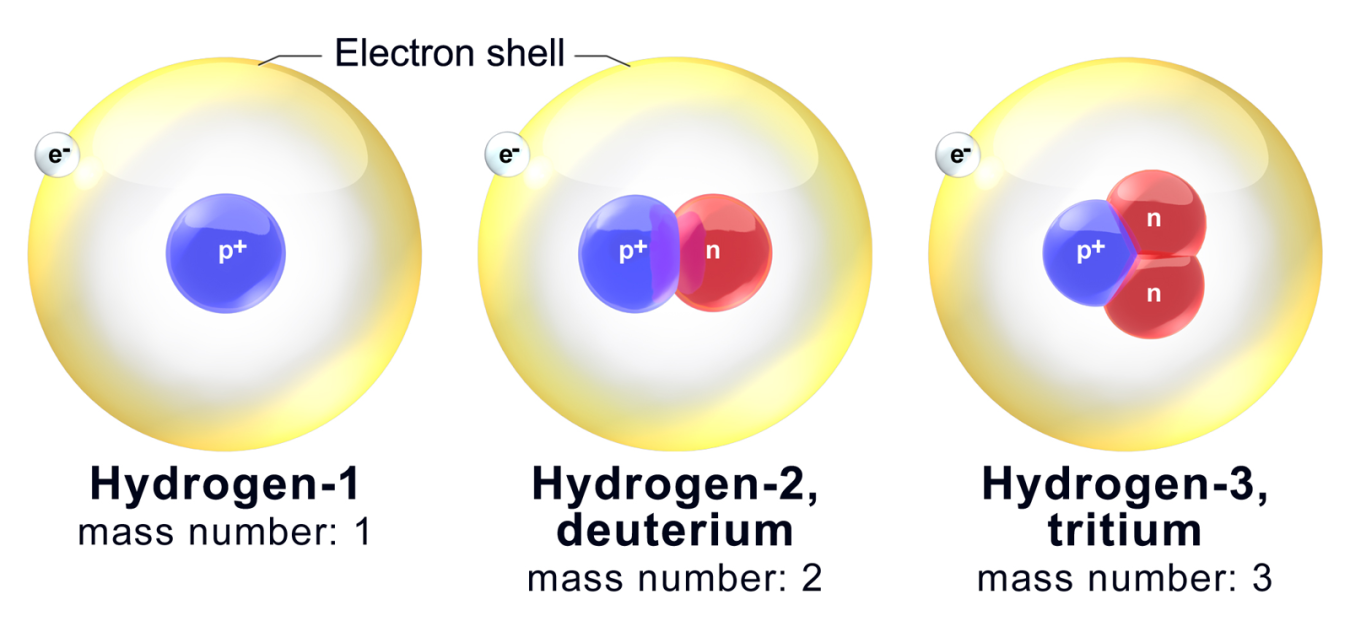
DOE and its predecessor organizations have been at the forefront of developing and producing unique isotopes for medical and basic science research for decades. Isotopes are different forms of the standard elements on the Periodic Table of the Elements. Certain radioisotopes (radioactive isotopes that decay) are extremely useful in medical research, imaging, and therapies. DOE has helped spur a renaissance in nuclear medicine. By providing rare and new isotopes, DOE has enabled doctors to use new treatments for metastasized cancers. DOE has also developed transformative technologies to reestablish stable isotope enrichment in the United States. In addition, it has led creative approaches for producing rare radioisotopes that are foundational to cancer imaging and therapy.
While research on protons and neutrons began at the accelerators and reactors of the DOE national laboratories to help us better understand the building blocks of the universe, this research has led to important tools now used in cancer diagnosis and therapies. Recent technologies that were developed for particle detectors have been adapted to identify tumors and monitor doses of radiation in patients.
DOE’s national laboratories have contributed to many other technologies and research results as well, including in protein folding and molecule production for chemotherapy drugs.
Highlights
-
-
- Biotechnology
- Cancer Research
- National Labs
- Explore Biology at DOE (Biology)
- Genomics
The President’s Cancer Moonshot aims to reduce the death rate from cancer by at least 50% over the next 25 years, and to significantly improve the lives of people, and their families, living with and surviving cancer. -
- Isotopes
- Biotechnology
- Cancer Research
- Nuclear Energy
- Next-Generation Energy Technologies
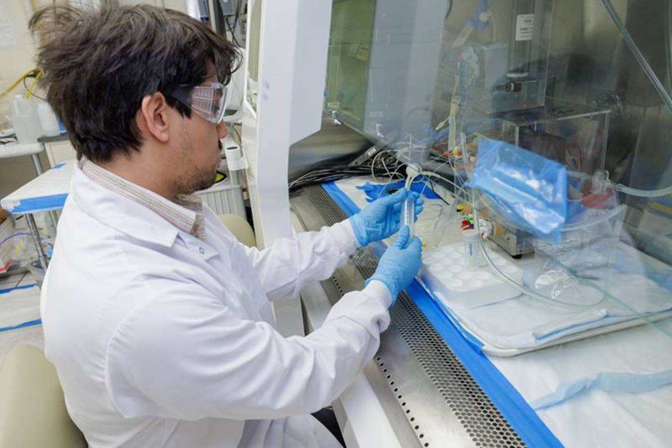
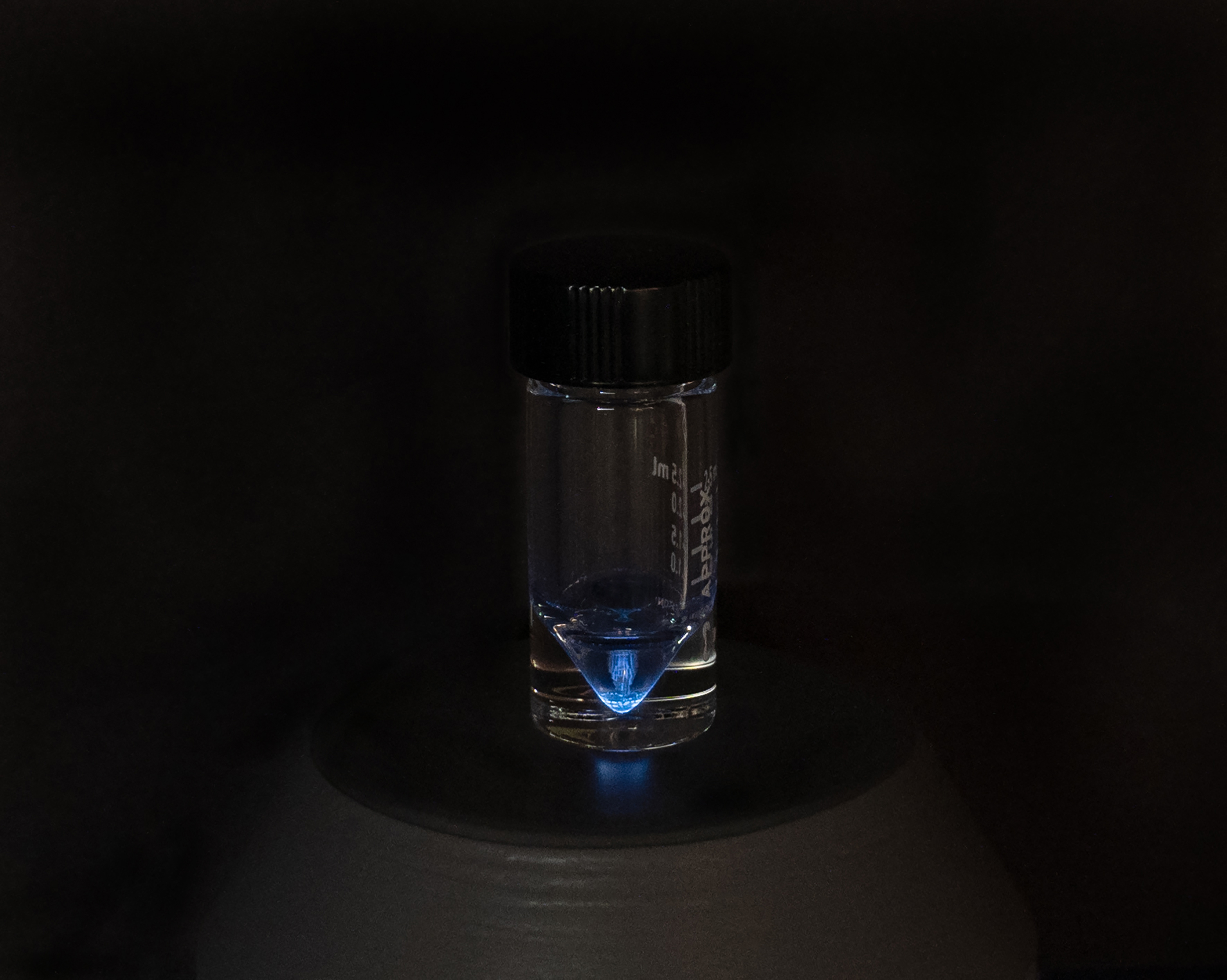
Researchers gain new insights into how the isotope astatine-211 interacts with resins commonly used to purify the isotope for therapeutic use.
Advances in cancer research enabled by DOE
DOE-supported tools and resources have enabled breakthroughs for cancer research in the past and will continue to do so in the future.
DOE’s supercomputers are playing a major role in cancer research. An algorithm developed by DOE’s Oak Ridge National Laboratory in partnership with the National Cancer Institute is speeding up classification of cancer pathology reports. The early results show that the algorithm dramatically reduces the time it takes for national cancer registries to process reports and the data to become available for public health monitoring. Another area where supercomputers have been essential has been modeling how cancer moves throughout the body. With the help of computers at the Oak Ridge Leadership Computing Facility and Argonne Leadership Computing Facility (both DOE Office of Science user facilities), researchers are using a full-body model of the human vascular system to track how cancer cells travel.
DOE-developed accelerator technology continues to advance the state of the art in external beam radiotherapy. Ultracompact accelerators that scientists first developed to make compact high energy physics accelerators have allowed accelerators to be made so small they can be mounted on an endoscope. Electronic brachytherapy delivers radiation directly to the tumor, which spares healthy tissues. Next-generation superconducting technologies developed for the Large Hadron Collider are being used to make beam delivery systems for bulky proton and ion accelerators more compact. This will enable more hospitals to afford these unique life-saving systems.
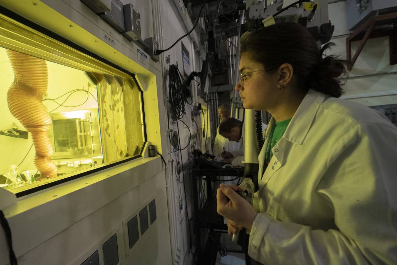
DOE produces many radioisotopes that are in short supply or available nowhere else and that are used in medical therapies. DOE also produces enriched stable isotopes that are used as starting materials for these radioisotopes. Some radioisotopes used in cancer therapy emit energetic particles that can cause a great deal of damage to a focused area, which is useful when targeting tumors. DOE produces two primary types of therapeutic radioisotopes: alpha particle emitters (e.g., actinium-225 and astatine-211) and beta particle emitters (e.g., rhenium-188 and yttrium-90). The introduction of alpha-emitters by DOE for the treatment of metastasized cancers has revolutionized cancer therapy treatments. Researchers from the DOE’s national laboratories and University Isotope Network have created new production processes to expand the supply of these radioisotopes. These new processes have enabled a number of active clinical drug trials.
DOE’s work enables private industry to enter mature isotope production markets. Medical imaging radioisotopes, like germanium-68 and strontium-82, allow physicians to identify tumors in people’s bodies in real time and see changes early on to better understand patient response. Both germanium-68 and strontium-82 were developed, and their markets were nurtured, by DOE. This action allowed DOE to transfer production to domestic private industry. These actions were in accordance with the DOE mission to act as a nonprofit governmental agency that does not compete with domestic private industry.
Nuclear physics research created the foundation for certain technologies that improve imaging and treatment monitoring. In breast cancer screening, nuclear medicine used alongside mammography reduces the number of false positive screening results. Based on research at DOE’s Jefferson Lab, scientists developed a tool to capture 3D molecular breast images at higher sensitivity. It provides up to six times better contrast of tumors, while maintaining the same or better image quality and halving patient radiation dose. Another cancer technology is based on material that scientists use to identify particles in experiments. Researchers have adapted it to monitor the radiation cancer patients receive in hard-to-reach areas.
Revisiting the CANcer Distributed Learning Environment (CANDLE) Project
This podcast from the DOE’s Exascale Computing Project focuses on the CANcer Distributed Learning Environment, or CANDLE subproject.
Press Releases
From Our Blogs
-

- Environmental and Legacy Management
- Public Health
- Cancer Research
- Biotechnology
- International Meetings and Forums
October 31, 2024 -
- Biotechnology
- Cancer Research
- Isotopes
- Next-Generation Energy Technologies
- Environmental and Legacy Management
October 8, 2024 -
- Biotechnology
- Cancer Research
- Genomics
- Explore Biology at DOE (Biology)
- Particle/High Energy Physics
September 19, 2024 -
- Biotechnology
- Research, Technology, and Economic Security
- Genomics
- Cancer Research
- Artificial Intelligence
August 21, 2024


